
What is a Posterior Tibial Tendon Dysfunction?
The posterior tibial tendon runs from the deep calf, where it attaches to the tibialis posterior muscle, passes behind the medial malleolus, the bony bump on the inner side of the ankle, and then attaches to the bones along the inner side of the foot. Its job is to help hold up the arch, support the foot during walking, and control the foot’s inward roll. A healthy tendon keeps the arch of the foot lifted. When it becomes irritated, stretched, degenerated, or torn, the arch may slowly drop, and the ankle may begin to roll inward.
Posterior tibial tendon dysfunction (PTTD) is the condition where the tendon that supports the inner arch of the foot becomes painful, weak, or unable to work properly. Posterior tibial tendon dysfunction is also called posterior tibial tendon insufficiency, or sometimes tibialis posterior tendon dysfunction. It is one of the most common causes of adult-acquired flatfoot.

Symptoms of Posterior Tibial Tendon Dysfunction
Posterior tibial tendon dysfunction usually causes pain or tenderness along the inner side of the ankle and foot.
Early symptoms often start as an aching or soreness behind the inner ankle bone or along the arch. The area may feel swollen or warm after walking, running, or standing for a long time. Some may notice foot pain when pushing off during walking, when climbing stairs, or when trying to rise onto the toes. The foot may feel tired, weak, or less stable than usual.
As the condition progresses, the arch may flatten, and the heel bone may drift outward. Shoes may wear unevenly, and the foot may appear wider or more collapsed than before. In later stages, pain can shift from the inside of the ankle to the outside of the foot and ankle as the joint alignment changes and other structures become compressed. This pattern is common when posterior tibial tendon insufficiency begins to affect the mechanics of the whole foot.
Causes of Posterior Tibial Tendon Dysfunction
Posterior tibial tendon dysfunction is usually caused by repeated overload, tendon degeneration, posterior tibial tendonitis, injury, or foot mechanics that place an extra strain on it.
It may become irritated after a sudden increase in walking, running, sports, or time on the feet. The structure can also weaken after a fall, repeated ankle rolls, acute trauma, ankle sprain, previous surgery, or long-standing overuse. Risk factors include flat feet, obesity, diabetes, high blood pressure, inflammatory arthritis, or previous foot and ankle injury. High-impact sport and poor footwear can also contribute, and over time, the tendon degenerates further.
Pes Planus and Posterior Tibial Tendon Dysfunction
Pes planus, or flatfeet, with posterior tibial tendon dysfunction means the arch has flattened as the tibialis posterior tendon is no longer supporting the foot well.
Some people are born with naturally flatter feet and never develop symptoms. In posterior tibial tendon dysfunction, however, the arch begins to collapse as it weakens. This is an acquired change, not simply a foot shape a person has always had.
When the arch drops because the posterior tibial tendon is not supporting the foot well, the whole foot can start to change position. The heel may roll outward, the front of the foot may turn outward, and the ankle may lean inward.
These changes can make walking less efficient and place extra strain on the ligaments, joints and tendons around the foot and ankle. In more advanced cases, the foot can become stiff and harder to correct without stronger support, such as a brace or surgery.
Posterior Tibial Tendon Dysfunction Stages
Posterior tibial tendon dysfunction stages describe how far the condition has progressed:
- Stage 1 usually involves pain, swelling, and inflamed tissue around the tendon, sometimes called posterior tibial tendonitis, without a fixed flatfoot deformity. The foot remains flexible, and the medial arch may still look fairly normal.
- Stage 2 involves a flexible flatfoot deformity. The arch begins to drop, but the foot can still be moved into a better position.
- Stage 3 involves a rigid flatfoot deformity, where the joints have become stiff and less correctable.
- Stage 4 involves the ankle joint as well, often with more advanced deformity and arthritis.
These stages are important because treatment varies as the condition progresses. Early stages are more likely to respond to orthoses, bracing, physical therapy and strengthening. Later stages may need more substantial support or surgical review if pain, deformity and loss of function are significant.
Diagnosis of Posterior Tibial Tendon Dysfunction
Posterior tibial tendon dysfunction is diagnosed through a physical examination, walking assessment and imaging tests when needed.
A physiotherapist’s examination usually includes checking the inner side of the ankle for tenderness and swelling, testing calf and foot strength, assessing arch height, and observing how the patient’s foot moves when standing and walking.
A common test is the single-leg heel raise. In early cases, this may be painful or difficult, causing pain along the inner ankle. In more advanced posterior tibial tendon insufficiency, the person may be unable to lift the heel well on the affected side. The clinician may also look from behind to see whether the heel is drifting outward or whether too many toes are visible from the outside of the foot.
X-rays can show flatfoot alignment and arthritis. Ultrasound scans may reveal thickening, fluid or tearing, and can identify a damaged posterior tibial tendon. MRIs can help assess the tendon quality, inflammation and associated soft tissues, ligament problems, midfoot arthritis, or a subtalar joint involvement. These imaging tests are useful when the diagnosis is unclear, symptoms are not improving, or surgery is being considered.
Posterior Tibial Tendon Dysfunction Treatment
Posterior tibial tendon dysfunction is usually treated first with load reduction, arch support, bracing, physical therapy and progressive strengthening.
Early treatment may include relative rest, ice after activity, nonsteroidal anti-inflammatory drugs when appropriate, and avoiding long walks, running, hills, or standing loads that flare up symptoms. Support is often important from the beginning. Orthoses can reduce the strain on the foot by supporting the arch and improving the foot position. A walking boot may be used for more painful cases to settle the symptoms before rehabilitation progresses, and a short course in a walking boot may be helpful for severe cases.
A posterior tibial tendon dysfunction brace may be recommended when the foot needs more support than a shoe insert can provide. A brace, and in some cases an ankle brace, can help relieve pain, reduce painful movement, support the medial arch, and improve confidence during walking. The type of posterior tibial tendon dysfunction brace depends on the stage of the condition, the foot’s flexibility, symptom severity, and activity needs.
Physiotherapy
Physiotherapy helps posterior tibial tendon dysfunction by improving the tendon load tolerance, foot control, calf strength, balance and walking mechanics.
A good plan begins by reducing the strain that keeps it irritated. This may mean modifying walking distance, changing footwear, using orthoses, or adding a brace for support. Once symptoms settle, the focus shifts toward rebuilding capacity. It needs enough load to adapt, but not so much that it flares up repeatedly.
Treatment may include manual therapy for stiff joints, techniques for the soft tissues of the calf and foot, gait retraining, balance work, and prescribed at-home exercises. Hip strength, calf muscle endurance, ankle mobility and foot control all affect how much demand is placed on the posterior tibial tendon and the surrounding soft tissues.
Adjunct Treatments
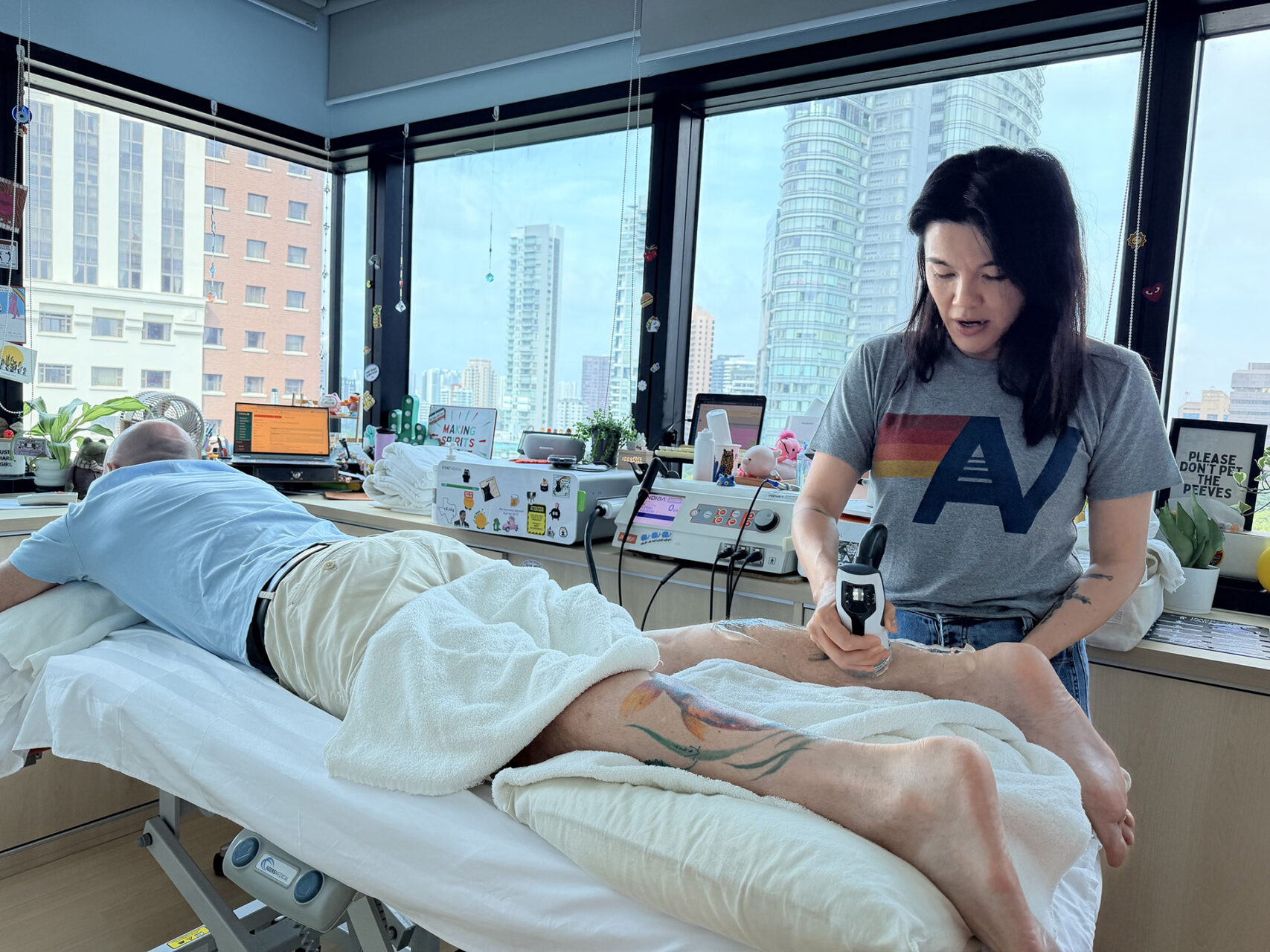
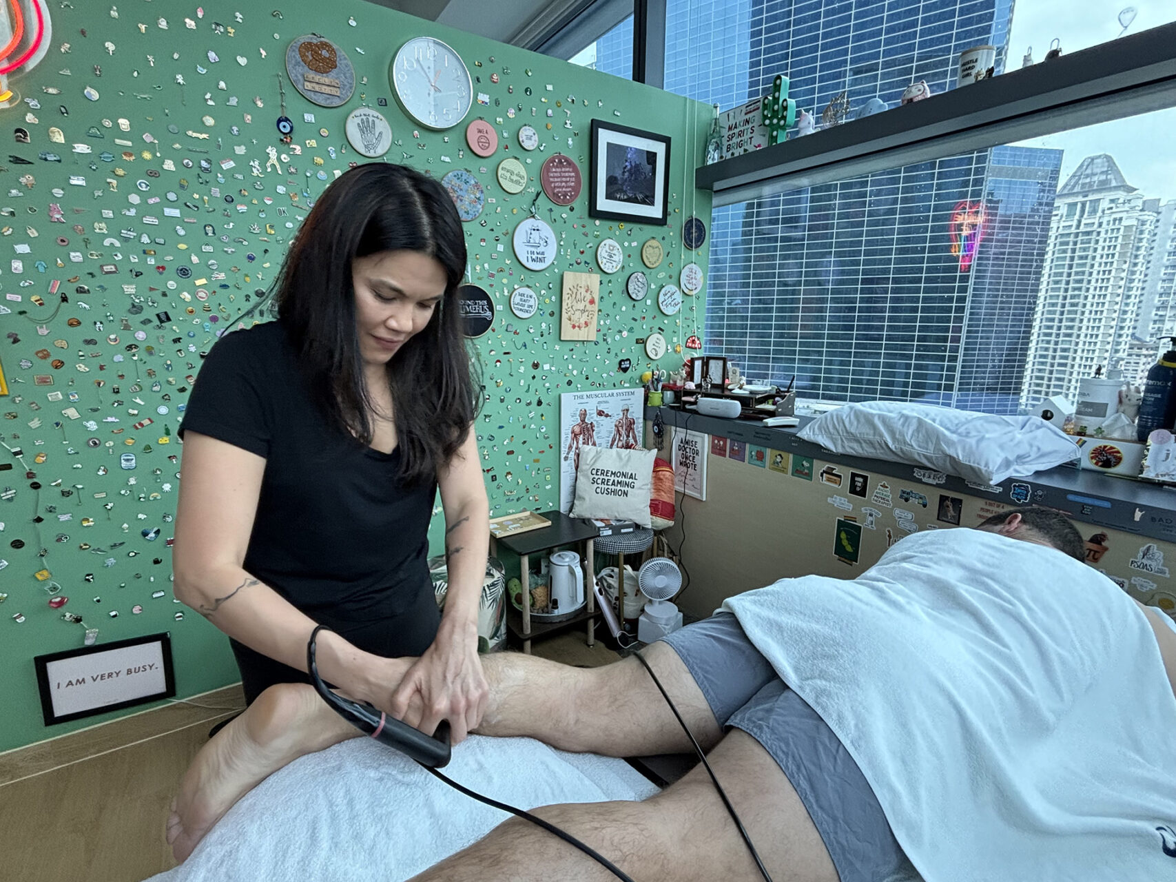
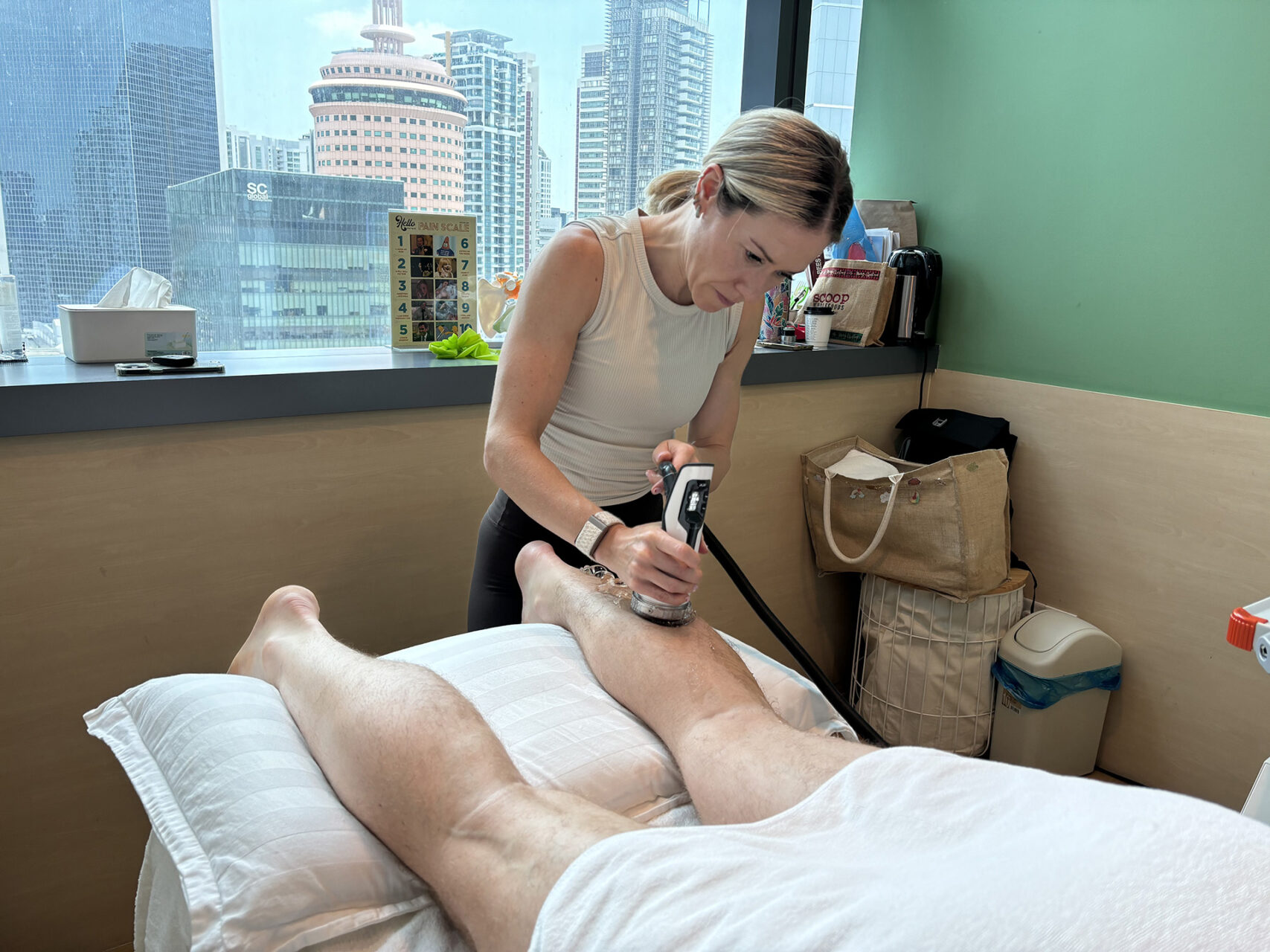
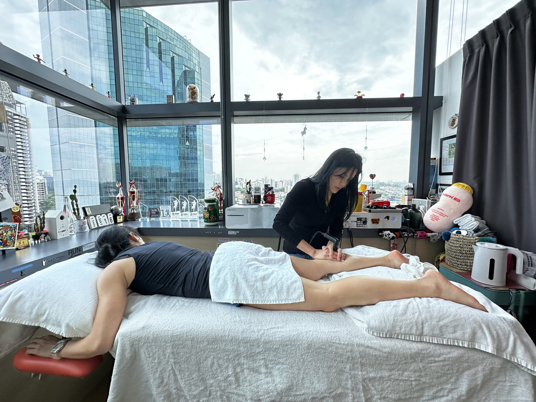
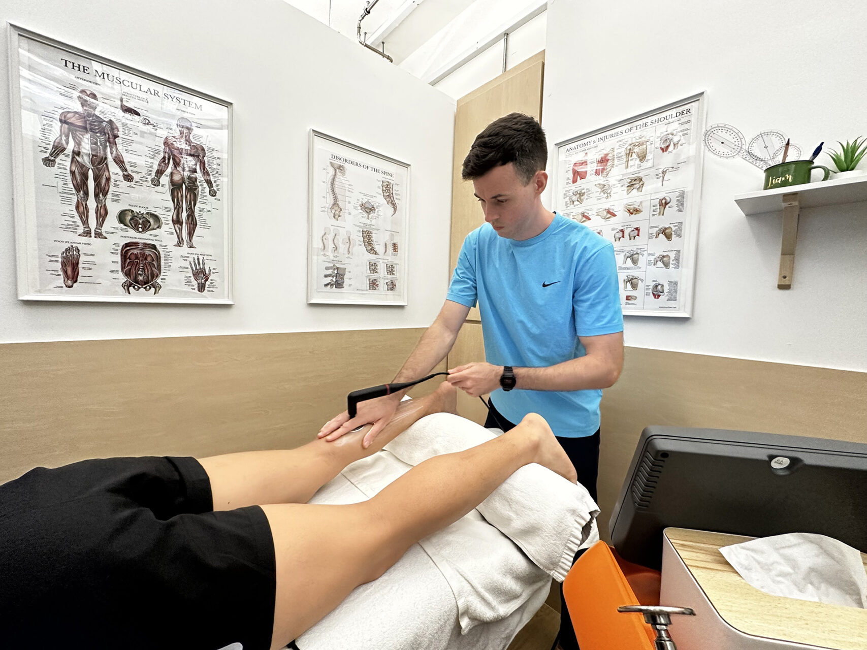
At HelloPhysio, adjunctive treatments may be used when clinically appropriate. INDIBA® may help with pain, stiffness, and movement tolerance as part of a broader rehabilitation plan. Dry needling may be considered if calf or foot muscle guarding is limiting movement, but it will not repair the guarding.
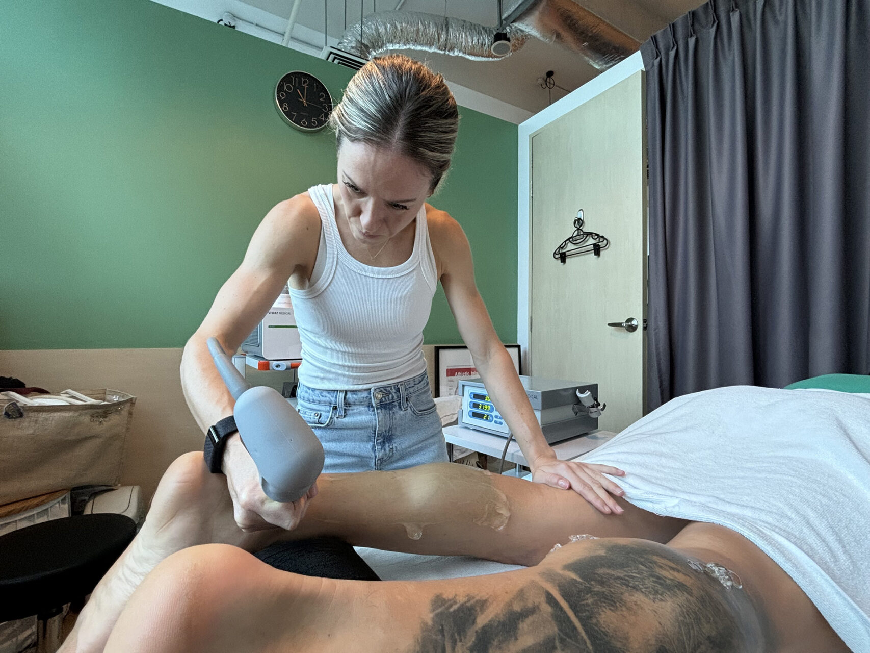
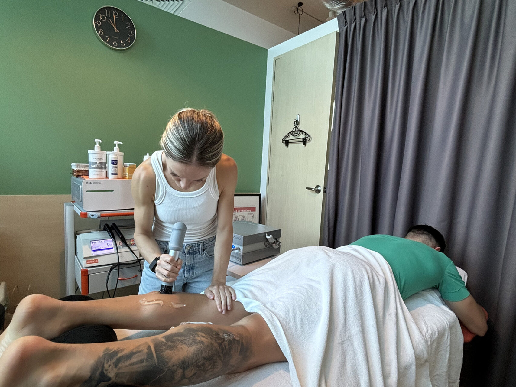
For chronic posterior tibial tendonitis that has not responded to standard care, radial Shockwave Therapy may be considered selectively, alongside a foot-and-ankle strengthening program.
Prescribed Exercises
Posterior tibial tendon dysfunction exercises should strengthen the tendon, foot, calf and hip muscles while protecting the arch from repeated overloads.
Early posterior tibial tendon dysfunction exercises may include gentle ankle movements, towel scrunches, arch setting, seated heel raises, and isometric inversion exercises. These help activate the muscles without placing too much of a strain on them. As symptoms improve, exercises may progress to standing calf raises, resisted inversion, balance work, step control and single-leg loading.
The exercise plan should be matched to the stage of the dysfunction. In early posterior tibial tendon dysfunction, strengthening exercises can often be progressed more actively. In a flexible flatfoot, arch support and bracing may need to remain in place while exercises build strength. In rigid deformities, exercise may focus more on maintaining mobility, reducing pain and improving function rather than restoring a normal arch shape.
Exercise should not cause sharp pain or a marked increase in swelling the next day. If it does, the load may need to be reduced. The aim is to keep the arch close to a normal arch position while building strength.
Surgery
Surgery may be needed when pain, deformity, or loss of function continues despite the appropriate nonsurgical treatment.
The type of surgery depends on the stage of posterior tibial tendon dysfunction. In earlier stages, procedures may aim to repair or support it and correct flexible deformity. This may include tendon transfer, often using the flexor digitorum longus tendon; bone realignment, such as a medial displacement calcanectomy; surgical reconstruction of the spring ligament; or procedures to support the arch. Achilles tendon lengthening or a gastrocnemius recession may also be needed when the calf is tight, and tendon transfer can be combined with these steps.
In more advanced rigid deformities, fusion procedures may be considered. Ankle fusion may be needed in severe cases when the ankle joint is involved. A foot and ankle surgeon experienced in this work can guide the discussion. After some procedures, a short leg cast is used to protect the healing.
Prevention of Posterior Tibial Tendon Dysfunction
Posterior tibial tendon dysfunction cannot always be prevented, but the risk can be reduced by supporting the arch, managing the training load, and strengthening the foot and ankle.
Prevention is especially important for people with flat feet, a history of ankle sprains, who engage in high-impact activity, or who spend long periods on their feet. Supportive shoes and proper footwear, gradual increases in running or walking volume, calf and foot strengthening, and early treatment of inner ankle pain can all help reduce overload.
For runners and active adults, the warning signs are usually clear before the condition becomes severe. Pain behind the inner ankle, arch fatigue, swelling, difficulty climbing stairs, or trouble with heel raises should not be pushed through for weeks. If pes planus with posterior tibial tendon dysfunction is already developing, early support and rehabilitation may help slow the progression and improve comfort.
How HelloPhysio Can Help
If inner ankle pain, arch collapse, or foot fatigue is affecting walking, running, or daily life, HelloPhysio can help. Our physiotherapists, working alongside a foot and ankle specialist, can assess your foot mechanics, identify the stage of your condition, and create a tailored treatment plan that includes support, strengthening exercises, and progressive rehabilitation. The best approach is usually a structured middle ground: reduce painful loads, support the arch, and rebuild strength gradually. Contact HelloPhysio to book a consultation today.
FAQs about Posterior Tibial Tendon Dysfunction
What does posterior tibial tendon dysfunction (PTTD) feel like?
It often feels like pain, swelling, or tenderness along the inside of the ankle and arch. The foot may feel weak or tired, and pushing off during walking or rising onto the toes may be painful or difficult.
What happens if PTTD is left untreated?
Untreated PTTD can progress to worsening tendon weakness, arch collapse, flatfoot deformity, stiffness, arthritis, and more difficulty walking. Early treatment is important because flexible changes are usually easier to manage than rigid deformity.
Is it okay to walk with PTTD?
Walking may be okay if symptoms are mild and settle afterwards. If walking causes limping, swelling, sharp pain, or next-day worsening, the tendon is likely being overloaded and needs more support or reduced activity.
When is surgery needed for PTTD?
Surgery may be needed when pain, deformity, or loss of function continues despite appropriate conservative care. It is more likely in advanced stages, rigid flatfoot, significant tendon failure, or cases involving ankle arthritis.
When is it too late for tendon surgery?
It may be too late for simple tendon repair when the foot has become rigid, the arch has collapsed significantly, or arthritis has developed. In these cases, surgery may still be possible, but it often involves realignment or fusion rather than tendon repair alone.
Can PTTD be cured?
Early posterior tibial tendon dysfunction often improves with support, physiotherapy, and strengthening. More advanced cases may not be fully “cured,” but symptoms, walking ability, and quality of life can often improve with the right treatment plan.