
What is a Medial Ankle Impingement?
The word “medial” means the inner side of the ankle, near the medial malleolus, which is the bony bump on the inside of the ankle. A medial ankle impingement, sometimes called a medial ankle impingement syndrome, is a painful pinching problem on the inner side of the ankle joint.
In simpler terms, the ankle runs out of smooth space. Instead of the joint gliding cleanly, tissue or bony growths get caught during an ankle dorsiflexion, which is the movement of bringing the foot upward toward the shin. This can make the ankle feel blocked, sore, swollen or unreliable.
Medial ankle impingement is less commonly discussed than other ankle impingement syndromes, but it can be just as limiting. Each pattern of ankle impingement syndrome, whether anterior ankle impingement, anteromedial impingement, posteromedial impingement, or posterior impingement, involves pinching at a different part of the joint capsule, articular surface, or surrounding soft tissues.
It often affects athletes and active adults who repeatedly load the ankle in deep positions, including runners, basketball and football players, gymnasts, dancers, and those with a history of ankle sprains. The condition may also develop after a fracture, an inversion ankle sprain, or a long-standing ankle instability.
Symptoms
A medial ankle impingement usually causes ankle pain, tenderness, and a pinching sensation along the inner front or inner side of the ankle.
Pain is often worse when the ankle is forced upward, such as during squatting, lunging, climbing stairs, running uphill, landing from a jump, or walking on uneven ground. Some people can point to one tender spot near the inner ankle joint line. Others describe a deeper ache that becomes sharper when the ankle is loaded.
Common symptoms of ankle impingement include swelling, stiffness, reduced ankle range of motion, clicking, catching, or a sensation that the ankle does not fully support your body weight.
In anteromedial impingement, pain is often intensified by lifting the foot up while turning it inward. Pain when pointing the foot downward usually suggests posterior ankle impingement rather than medial ankle impingement. This happens when the back of the ankle joint gets pinched during movements such as pointing the toes, jumping, kicking, or pushing off. The pain is often linked to irritation of the back of the talus, one of the main ankle bones.
Symptoms can be easy to mistake for a sprain that has not fully healed. This is especially true when pain begins after an ankle roll. If the inside of the ankle remains tender, swollen, or blocked weeks after the injury, a medial ankle impingement should be considered alongside other forms of ankle impingement that may produce overlapping anterior or chronic ankle pain.
Causes
Medial ankle impingement syndrome is usually caused by repeated ankle injury, repetitive loading, bony spurs, scar tissue, or instability.
A common pattern starts with ankle trauma. After an ankle sprain or fracture, the joint may heal with soft tissue scarring or mild changes in alignment. If the ankle remains stiff or unstable, the inner joint can be repeatedly compressed. Over time, the body may form osteophytes, called bone spurs. These bone spurs can form on the inner ankle bones and start to bump into each other when the ankle bends upward. This pinching can irritate the joint and cause pain, stiffness, and a blocked feeling in the ankle.
Foot shape can also contribute. A subtle high-arched foot, called a cavus foot, may shift the load toward the inside or outside of the ankle, increasing joint stress. Repeated ankle sprains, poor ankle control, and chronic ankle instability can make this worse. Tightness in the calf muscle can also limit how far the ankle bends upward. This can place extra stress on the front or inner part of the ankle, contributing to ongoing pain. Medial ankle pain after repeated sports activity, running, or prior ankle sprains is a common reason patients seek care at sports physiotherapy clinics.
The condition can also be linked with other problems inside the ankle, such as osteochondral lesions of the talus, loose bodies, stress fractures, or early medial ankle joint wear. Related problems can also develop after ankle sprains or ligament injuries. Scar tissue may build up near injured ligaments after the ankle rolls inward, causing pinching on the outer side of the ankle. Injuries to the deltoid ligament on the inner side of the ankle can lead to pain and pinching at the back and inner part of the joint. Repeated stress through the ankle joint over time can also cause ongoing irritation and impingement.
Diagnosis
Medial ankle impingement is diagnosed through a physical examination and, when needed, imaging.
A physiotherapist will ask how the pain started, where it is felt, what movements provoke it, and whether there has been a previous sprain, fracture, or repeated ankle rolling. The physical examination usually includes a palpation over the inner ankle and medial malleolus, range-of-motion testing, strength checks, balance assessment, gait observation, and tests for ankle instability.
The painful area is often tender to the touch. Symptoms may be reproduced by dorsiflexing the ankle, especially when the foot is also turned inward. The clinician will also check for other causes of medial ankle pain, such as posterior tibial tendon irritation, tarsal tunnel syndrome, medial malleolar stress injury, subtalar joint problems, or arthritis affecting this synovial joint.
X-rays can show bone spurs, joint narrowing, or changes in the ankle joint. Sometimes special X-ray angles are needed because small spurs, especially on the inner side of the ankle, can be missed on standard views.
CT scans give a more detailed picture of the bones and can show the size and shape of spurs or loose bone fragments. An MRI is useful when the problem may involve soft tissues, cartilage, joint swelling, ligament injury, or hidden bone bruising.
In simple terms, X-rays and CT scans show the bones well, while an MRI provides more detail on the soft tissues around the ankle, including ligaments, tendons, scar tissue, inflammation, and cartilage damage. This helps identify whether the pain is coming from bone spurs, trapped scar tissue, ligament thickening, swelling, or it is another ankle problem.
Medial Ankle Impingement Treatment
Treatment for medial ankle impingement syndrome usually begins with reducing the irritation, improving ankle mechanics, and restoring strength and control.
Early Care
Early care may include a short period of activity modification, reducing painful squats, hills, jumping, or cutting drills. Ice, compression, and anti-inflammatory medication may be used when appropriate to reduce the pain and swelling. In more irritated cases, an ankle brace, supportive taping, or a short period in a walking boot may help protect the joint while symptoms settle.
Physiotherapy
Physiotherapy helps medial ankle impingement by improving your movement, reducing the overload, and rebuilding the strength and control that protect the joint.
The first goal is to understand why the inner ankle is being pinched. For one person, the main issue may be stiffness after a sprain. For others, it may be poor balance, weak calf strength, altered running mechanics, or repeated ankle instability.
Manual therapy may be used to improve joint mobility and reduce stiffness in the tibiotalar and subtalar joints, the foot, and the calf. Soft tissue treatment may help if the calf, tibialis posterior, Achilles tendon, or other lower-leg muscles are guarding around the painful joint.
Dry needling may be considered when muscle tightness or trigger points are limiting movement, but it does not remove bone spurs or directly treat the articular surface of the joint.
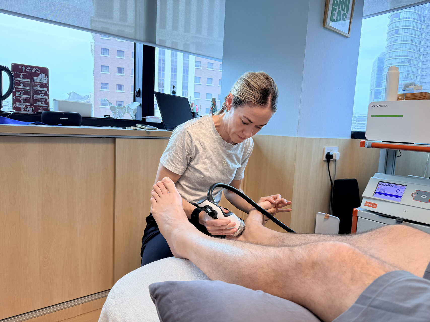
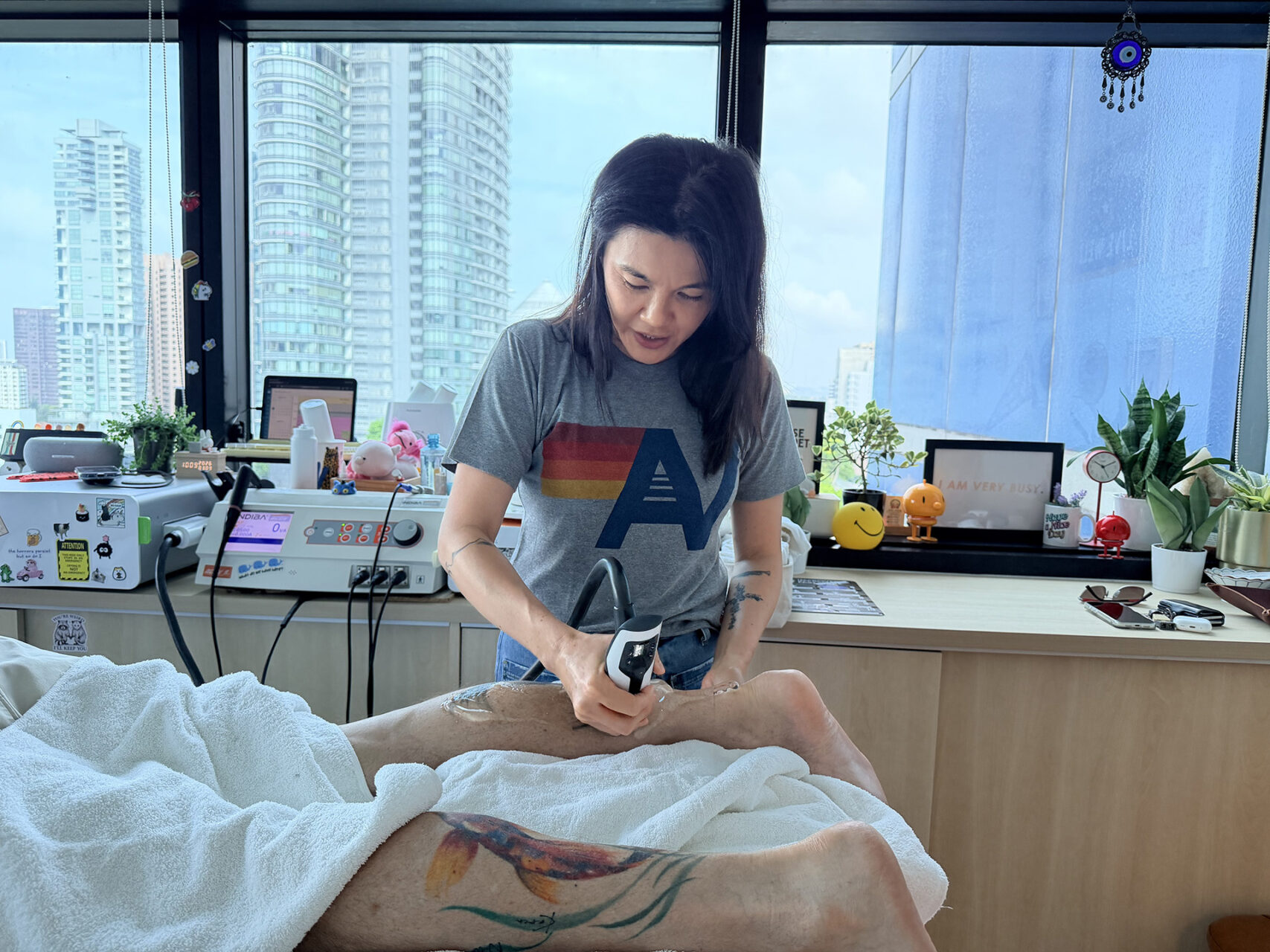
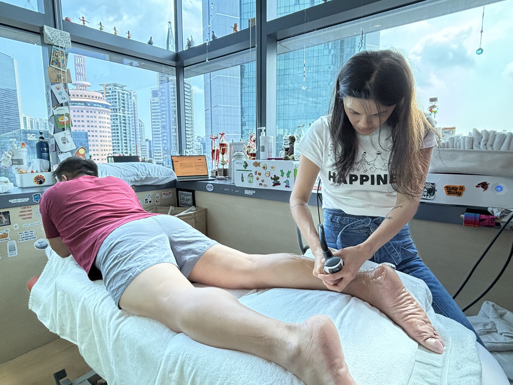
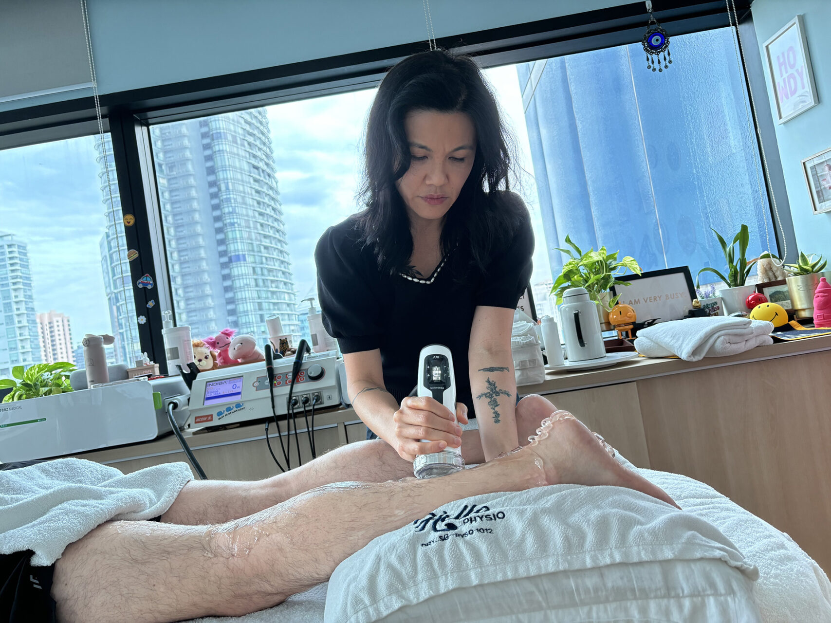
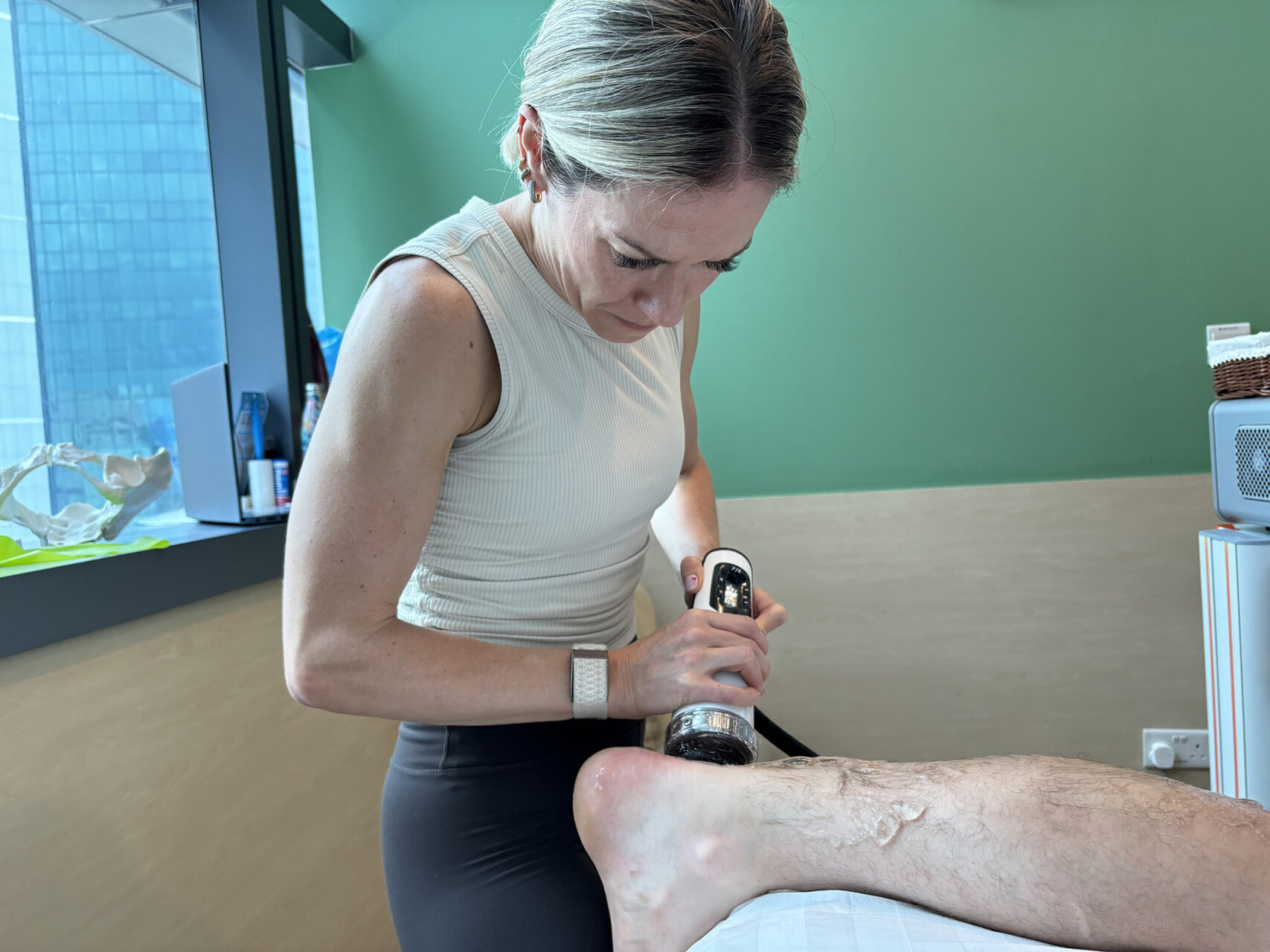
INDIBA® may be used as an adjunct to support pain relief, tissue recovery, and mobility when symptoms are irritable.
Prescribed Exercises
Exercises for medial ankle impingement should improve ankle mobility, calf strength, foot control, and balance without forcing the painful pinch.
Early exercises often focus on gentle movement. Ankle pumps, circles, and controlled weight shifts can help reduce stiffness. Calf stretching may be useful if tightness is limiting dorsiflexion, but stretching should not be pushed to the point of sharp inner ankle pain.
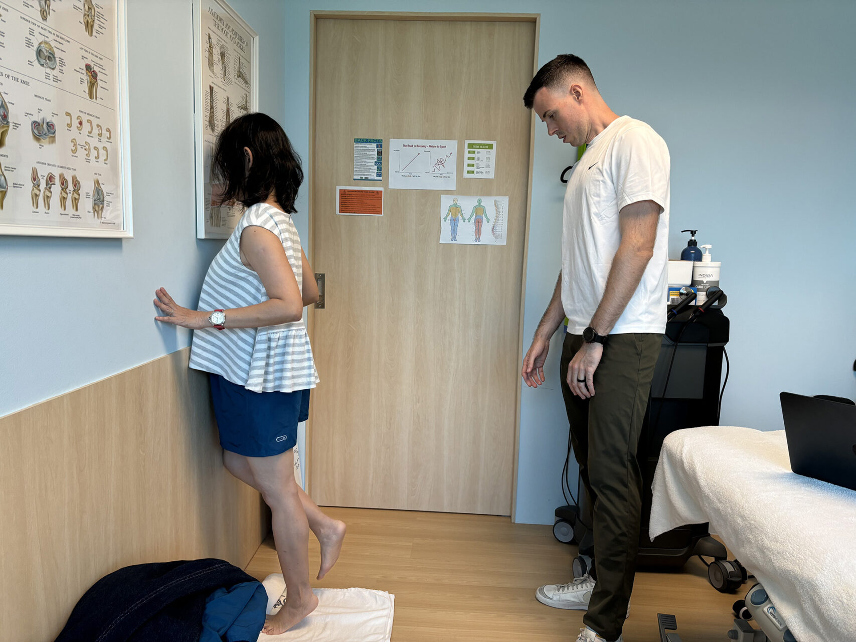
As symptoms settle, strengthening becomes more important. Supported calf raises, seated heel raises, resistance-band inversion and eversion, and controlled step work can help the ankle tolerate load. Balance exercises, such as single-leg stances near a stable surface, help retrain proprioception. Proprioception is the body’s sense of joint position, and it is often reduced after ankle sprains.
For active patients, exercises should eventually resemble the demands of sport. A runner may need graded hill tolerance and single-leg calf endurance. A court-sport athlete may need control over landing, pivoting, and change of direction. The right exercise plan should build capacity without repeatedly reproducing the feeling of being blocked.
Ankle Brace
If instability is present, an ankle brace can be helpful. If the foot shape is contributing, orthotics may be recommended to improve the load distribution. In those with a subtly high-arched foot, orthotics may help reduce abnormal loading through the medial ankle. Intensive exercise that repeatedly provokes the painful pinch may worsen symptoms, so exercise must be graded carefully.
Corticosteroid Injections & Surgery
Corticosteroid injections may be considered if inflamed soft tissues are a major contributor and pain is stopping progress. Surgical intervention is considered when symptoms persist despite conservative care, especially when a bone spur, loose body, or a clear mechanical block is present.
Surgical treatment may involve open or arthroscopic removal of the offending bone spur or inflamed tissue. Keyhole surgery, called an arthroscopy, is often used to treat ankle impingement. It can help remove scar tissue, inflamed tissue, or bone spurs at the front, side, or back of the ankle that developed after sprains, repeated strain, or irritation around extra bone structures near the ankle joint. Some patients may also benefit from an ankle brace after surgical intervention while the soft tissues heal.
Recovery
Recovery from medial ankle impingement syndrome depends on whether the problem is primarily soft-tissue irritation, instability, or a bony mechanical block.
Milder soft tissue cases may improve over several weeks with good load management and physical therapy. More chronic cases can take a few months, especially if there has been a repeated sprain, longstanding stiffness, or poor ankle control. If bone spurs are large and continue to abut during movement, conservative care may reduce symptoms but not fully remove the mechanical limitation.
Surgical intervention may be considered when symptoms persist after a reasonable period of conservative treatment. Rehabilitation is still important after surgery because the ankle must regain motion, strength, control of swelling, and confidence before full return to sport or impact activity. After surgical removal of significant bone, jumping activities may be delayed for several weeks while the bone and soft tissues settle.
Prevention
Medial ankle impingement syndrome may be prevented or reduced by fully rehabilitating ankle injuries and avoiding repeated overloads through a stiff or unstable joint.
The most important step is not ignoring ankle sprains. Pain may improve before strength, balance, and joint control have fully returned. If the ankle keeps rolling, swelling, or feeling blocked, it needs proper rehabilitation. Supportive footwear, progressive strengthening, balance training, and a gradual return to running or sport can reduce the chance of ongoing joint irritation.
Patients with high arches, recurrent ankle sprains, or instability may benefit from footwear review, orthotics, taping, or an ankle brace during higher-risk activity. These supports are most useful when they help the person move well while strength and control are rebuilt.
How HelloPhysio Can Help
If inner ankle pain is limiting your ability to walk, run, use stairs, or participate in sports, HelloPhysio can help. Our physiotherapists will assess the joint, identify the likely driver of your symptoms, and create a treatment plan that supports pain relief, mobility, strength, and a safer return to activity. Contact HelloPhysio to book a consultation.